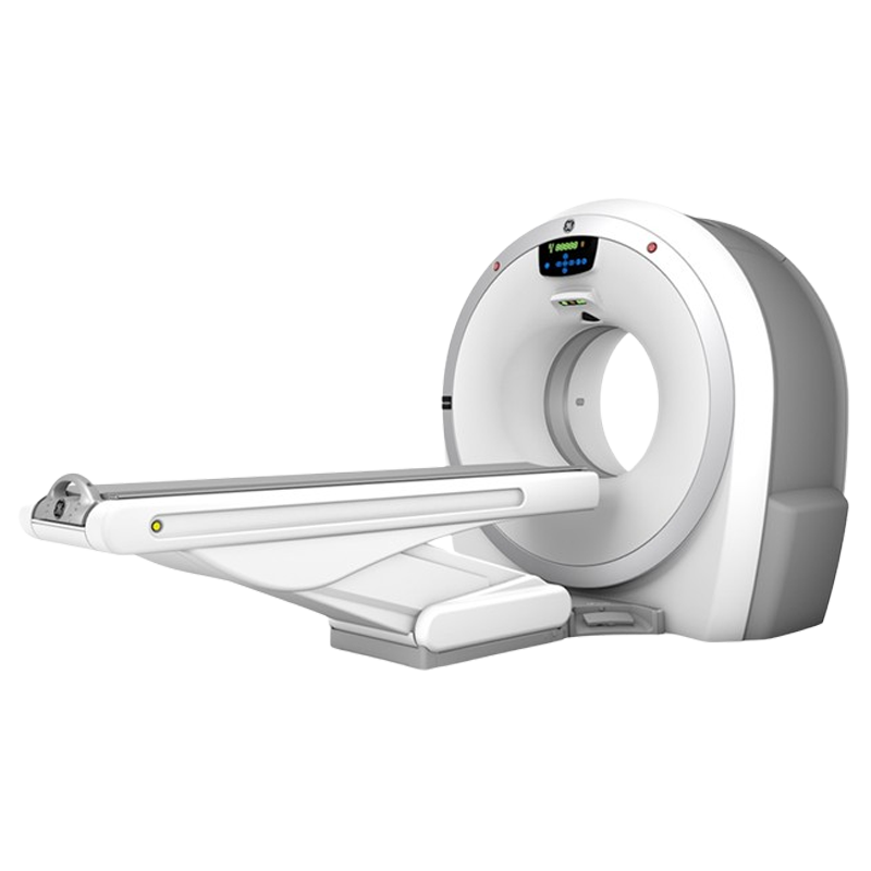
Features
Revolution ACT allows you to not only provide better patient care, but also provide it to more patients. Avant-garde technologies help you feel more confident about moving to the next level – clinically and economically. Designed for consistent high-quality results, Revolution ACT enables caregivers make the most informed diagnoses possible.
Higher Performance
- Clarity Panel Detector: Innovative detector technology that delivers exceptional spatial resolution
- Lower Electronic Noise: Integrated detector design built with modern chipsets and DAS on Detector (DoD) for high signal-to-noise performance
- Efficient Design: Compact and power-efficient design with intelligent thermal management for quicker scans
- Ultra Kernel: Adaptive Enhance Level Adjustment (AELA) can improve visual spatial resolution while maintaining pixel noise standard deviation and artifact
Smart Dose
- ASiR*: Provides breakthrough image quality in multi-slice CT exams
- ViSR*:Delivers up to 20% improvement in image quality at the same dose. Helps manage photon starvation in large-size patients and wide/dense anatomical objects.
- ODM: Reduces radiation dose by up to 40%. Acts as virtual shield to improve image quality
- 3D mA Modulation: Enables the system to optimize tube current in x-y-z directions with negligible effect on image quality
- Dose Watch: Tracks and monitors patients’ cumulative radiation dose over time and takes intelligent steps to prevent excessive radiation dose

Smart Flow Technologies
- Digital Tilt: Simple and fast digital tilt enables more effective scanning and ups workflow by 28%. Helps manage challenging and less cooperative patients
- IQ Enhance: Advanced algorithms enable helical pitch acceleration up to 2.7x and helps patients breathe easier with shorter breath hold time


Technology
Advanced applications of Revolution ACT help you take your practice to the next level. It provides you with the necessary information to make the best possible diagnoses for patients.
- Volume viewer: Get more information on the spatial relationships of different structures. Make 3D visualizations routine
- Vascular imaging and processing: View oblique cross-sections of vascular images and rotate curved views to more clearly visualize vascular lesions
- Navigation and fly-through: Use Virtual Endoscopy to visualize intra-luminal structures. A virtual ‘fly-through’ mode lets you view images dynamically
SmartPrep with dynamic transition: Transition from monitor phase to scan phase automatically


